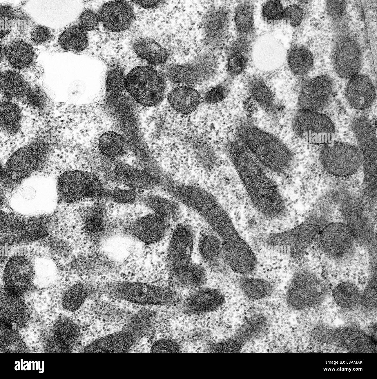
Mitochondria Micrograph
May 06, 2024
Images for Mitochondria Micrograph
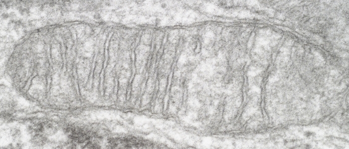
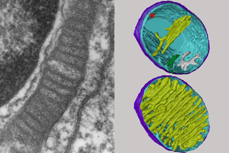
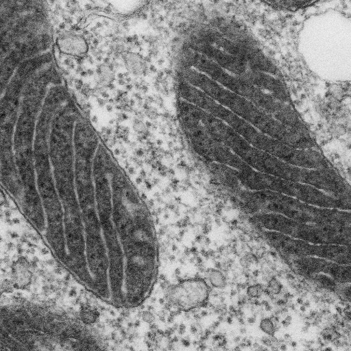
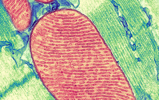
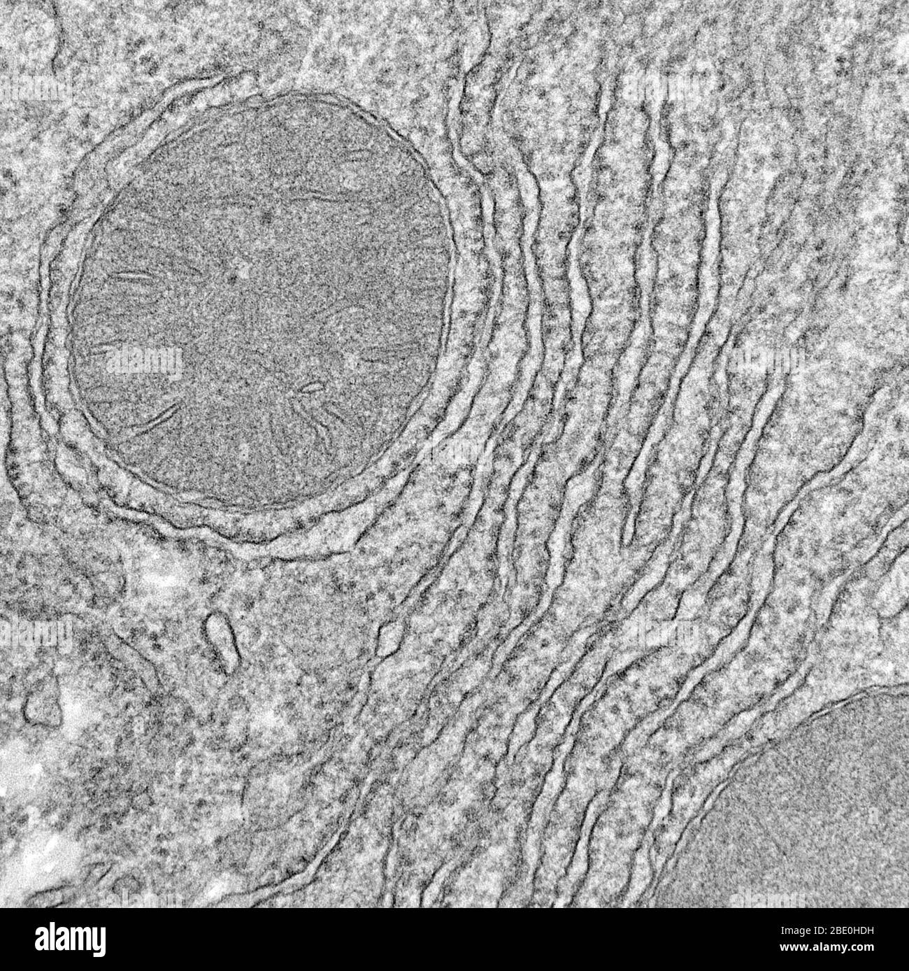
Medical School — Scanning electron micrograph of the mitochondria
ATP – Structure & Function/ Anaerobic and Aerobic Respiration
Evolution of the Mitochondrial Genome
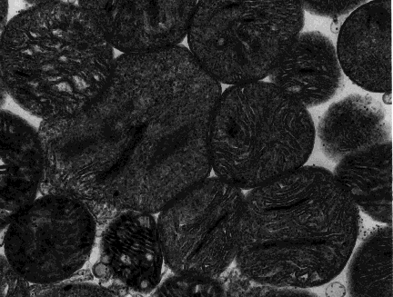
TEM micrographs of mitochondria of Micrasterias before and during
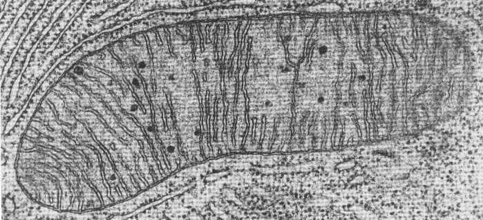
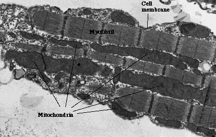
A mitochondria as seen with an electron microscope. Because you look at
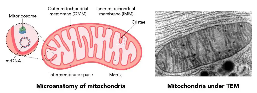
Mitochondria | Contexo.Info
Picture
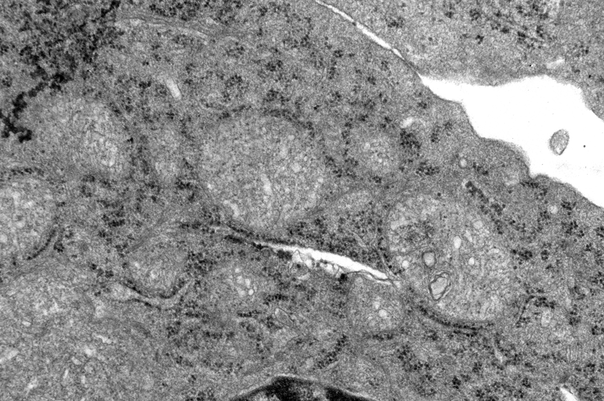
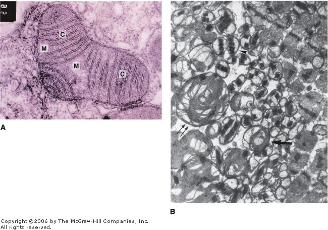
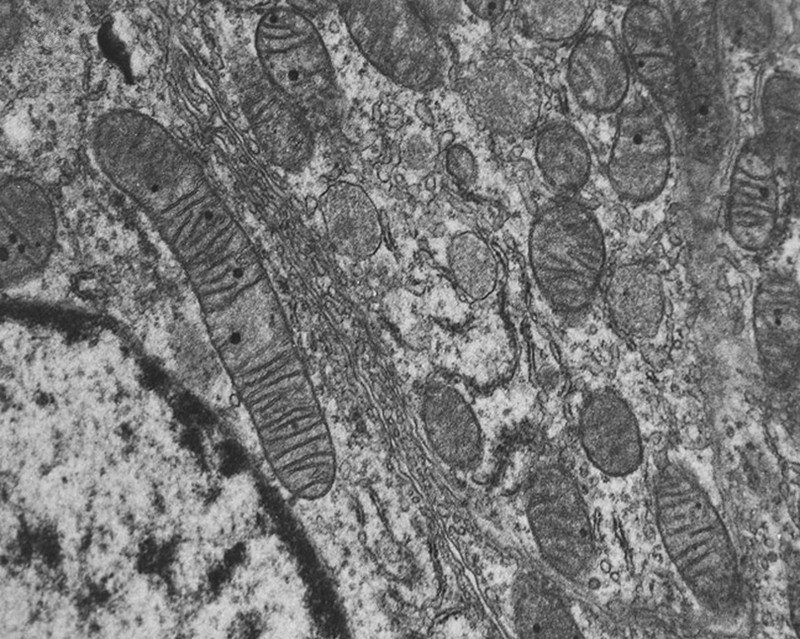
Electron micrographs of mitochondria (M). A: Mitochondria (M) had
Electron micrograph of mitochondria in Tebufenozide treated last instar
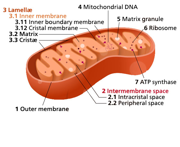
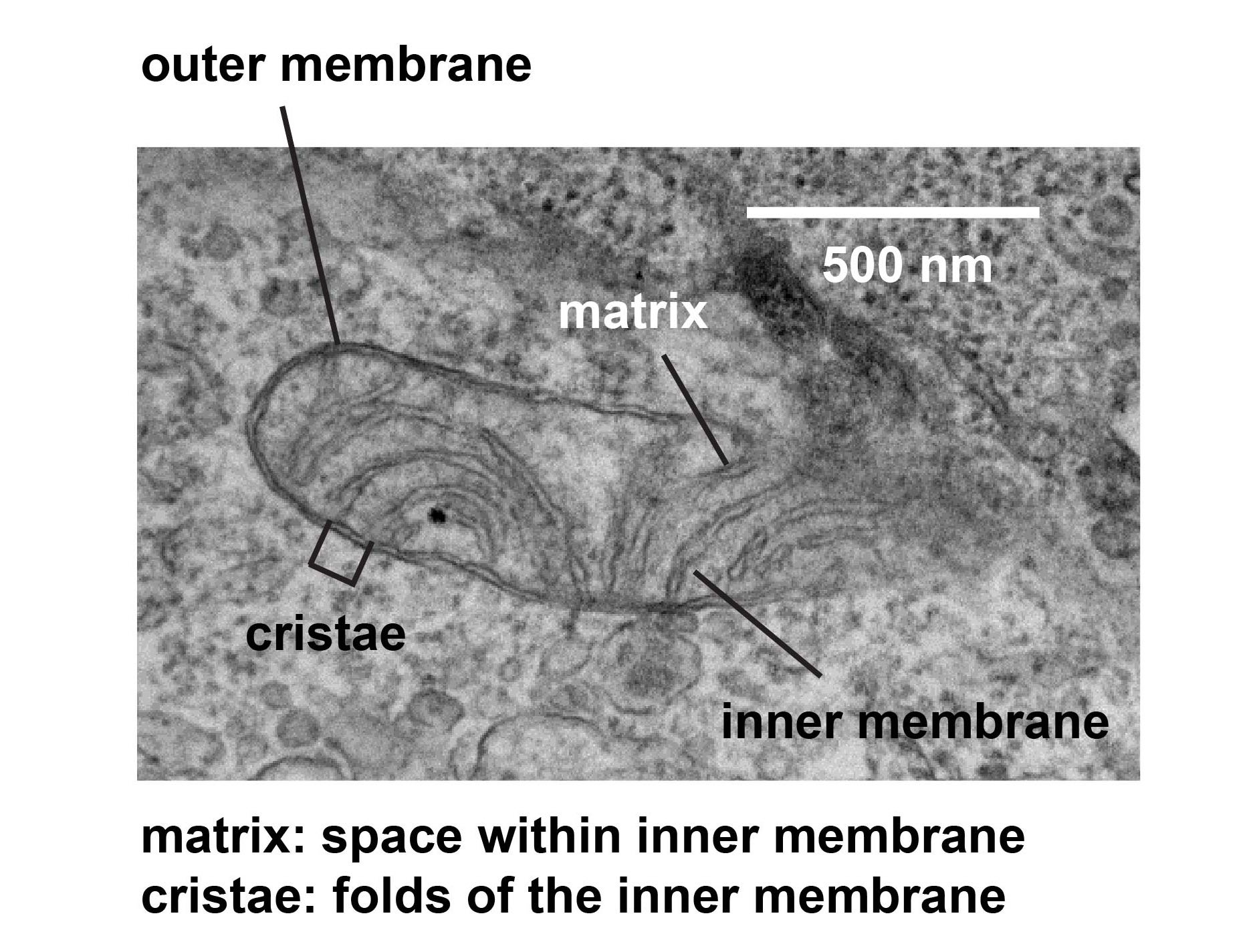
Mitochondria Microscopy detailed labelled 3D model | CGTrader
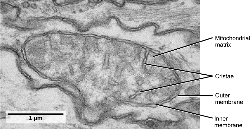
5.12: Mitochondria and Chloroplasts - Biology LibreTexts
Electron micrograph of a mitochondrion in a cell of the bat pancreas
Unit 1 - Biology 12 Textbook
Electron microscopy. (A) Normal mitochondria morphology was observed in
Stage 23. a Electron micrograph of photoreceptors. Mitochondria (m) in
Mitochondrion electron micrograph — Science Learning Hub
Ribosomes, Mitochondria, and Peroxisomes | Biology for Majors I
Electron micrographs of mitochondria from different tissues
Representative electron micrographs of mitochondrial alterations in
Mitochondria under the microscope — Science Learning Hub
1.7 Mitochondria – Plant Anatomy and Physiology
Electron micrographs of mitochondria isolated from C. fascicuiatu. (a
However, as shown in the fluorescent micrograph below, each cell
Mitochondrial Unfolded Protein Response | Germain Lab
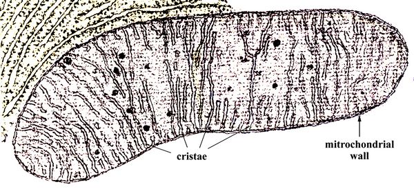
mitochondrion showing cristae (SEM)
A roadmap for metabolic reprogramming of aging « Kurzweil
Giant Mitochondria in the Myocardium of a Patient With Mitochondrial
De Histology: Mitochondria
mitochondria - Share Healthy Life
Marvelous Mitochondria
Electron microscopic image of isolated mitochondria. Their shape and
Electron micrograph of giant supercoiled DNA from mitochondria of(6
Mitochondria - the powerhouses of the cell - definition, structure
What Living Things You Can See Under a Light Microscope? - Rs' Science
Trasmission electron micrographs of mitochondria from cauliflower and
Mitochondria structure
Figure 3 from Recent structural insight into mitochondria gained by
Electron micrographs of mitochondria in left ventricular myocardial
This transmission electron microscope image reveals mitochondria in a
Biochemistry, University of Toronto – G. Angus McQuibban
Transmission Electron Micrograph Of Mitochondria Photograph by Dr Gopal
3.4 Unique Characteristics of Eukaryotic Cells – Microbiology: Canadian
Transmission electron microscopy of mitochondria in the testicular
Enzyme Restores Function with Diabetic Kidney Disease
A. Electron micrographs of mitochondria at different times (hour
Mitochondria Micrograph High Resolution Stock Photography and Images
Mitochondria Stock Photos - Download 189 Images
Mitochondria Micrograph Stock Photos & Mitochondria Micrograph Stock
Quia - Cell Parts and Functions Flash Cards
Electron micrograph of abnormal mitochondria in the right biceps
Gallery2 - Keele University
Electron microscopy of RV mitochondria. Electron microscopy of
(PDF) Unusual mitochondria of the bovine oocyte
—Mitochondrial transmission electron microscopy (TEM) from lung rat
| Electron microscopy indicated abnormal mitochondria. (A) Normal
Numbers and morphology of mitochondria in CSCs and non-stem cancer
‘Social’ Mitochondria, Whispering Between Cells, Influence Health
Edexcel IAL Biology
Abnormal mitochondria in mouse and human PKD cells and tissues. (a
Transmission electron micrograph of oocyte mitochondria at dynamic
Cell Membrane Micrograph High Resolution Stock Photography and Images
Observation of mitochondria with transmission electron microscopy
Electron micrograph, showing cells containing numerous mitochondria
Electron micrograph of mitochondrial tethering by truncated Mfn1. Scale
Electron micrographs of mitochondrial human mononuclear cells treated
Transmission electron micrographs of mitochondrial and axonal
Mitochondria Dr.Jastrow's EM-Atlas
Pictures of isolated mitochondria by transmission electron microscopy
Mitochondria structure
Ribosomes Under Electron Microscope - Micropedia
Mitochondrial ultrastructure in YtA and YtB. Transmission electron
Pin on Mitochondria
Mitochondria | Science cells, High school biology, Molecular biology
Mitochondria stock image. Image of bacteria, energy, mitochondria
Transmission Electron Micrograph Of Mitochondria Photograph by Dr Gopal
Blog do Enem: simplificado como deve ser ~ Sandyt Agregador
Mitochondrial ultrastructure and subcompartments. Electron micrograph
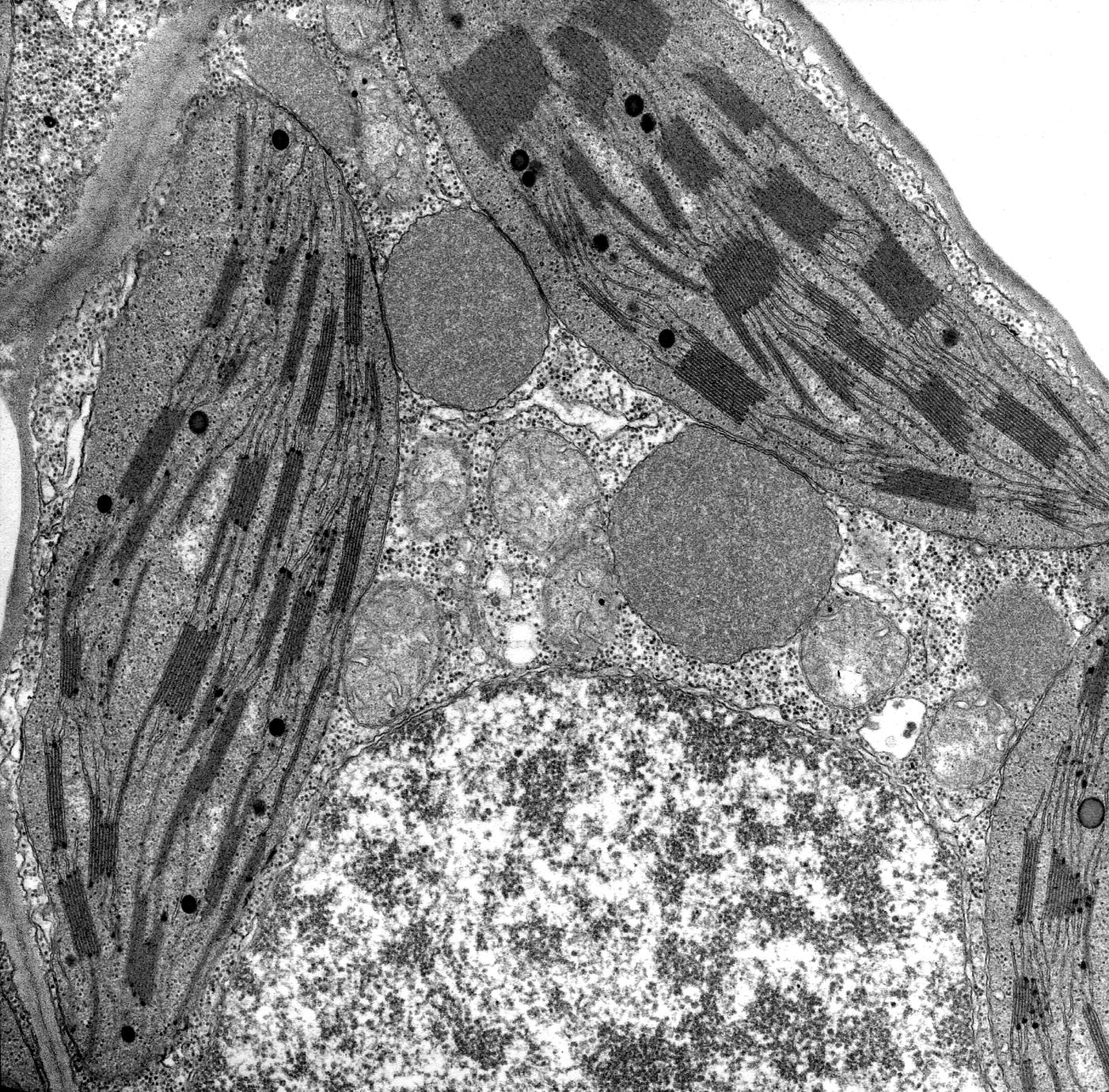
Nucleus, glyoxisomes, chloroplasts, and mitochondria - magnification
Morphological alterations of DMD mitochondria. A: Transmission electron
Transmission Electron Micrograph Of Mitochondria Photograph by Dr Gopal
A new spin on energy independence | Broad Institute
Pictures of isolated mitochondria by transmission electron microscopy
Representative electron photomicrographs of mitochondrial | Download
Mitochondrion, TEM - Stock Image - C023/1020 - Science Photo Library
Transmission electron micrographs of benthic foraminiferal
Transmission electron microscopy analysis of cell body mitochondria
Mitochondria appearance under electron microscope (EM × 6000); A
Electron micrograph of mitochondria in liver cell - Stock Image - G465
Mitochondria Micrograph High Resolution Stock Photography and Images
Case 1. On electron microscopy, (4A) mitochondria (arrows) (10,000x
CC BY-NC 4.0 Licence, ✓ Free for personal use, ✓ Attribution not required, ✓ Unlimited download
Free download Medical School Scanning electron micrograph of the mitochondria,
ATP Structure Function Anaerobic and Aerobic Respiration,
Evolution of the Mitochondrial Genome,
TEM micrographs of mitochondria of Micrasterias before and during,
A mitochondria as seen with an electron microscope Because you look at,
Mitochondria ContexoInfo,
Picture,
Electron micrographs of mitochondria M A Mitochondria M had,
Electron micrograph of mitochondria in Tebufenozide treated last instar,
Mitochondria Microscopy detailed labelled 3D model CGTrader,
512 Mitochondria and Chloroplasts Biology LibreTexts,
. Additionally, you can browse for other images from related tags. Available online photo editor before downloading.
Mitochondria Micrograph Suggestions
Mitochondria Micrograph links
Keyword examples:
Site feed


